2026,
13(8):
1826-1841.
doi: 10.1109/JAS.2025.125705
Abstract:
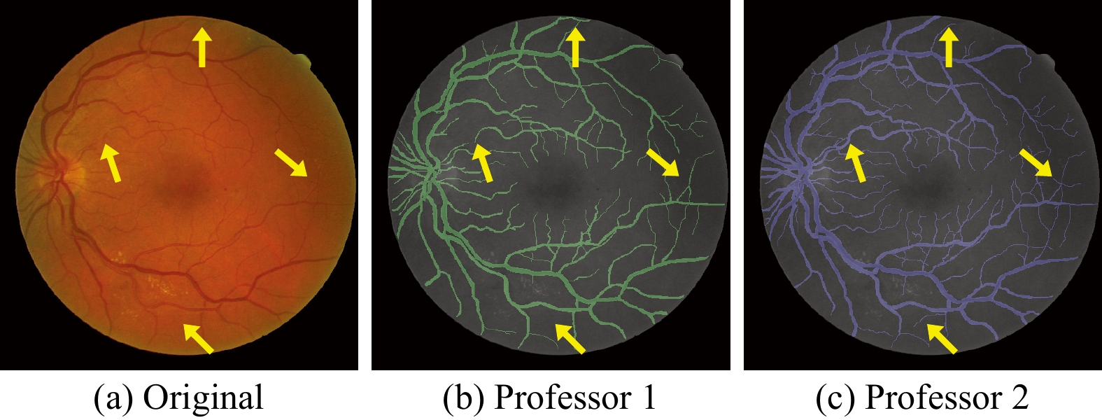
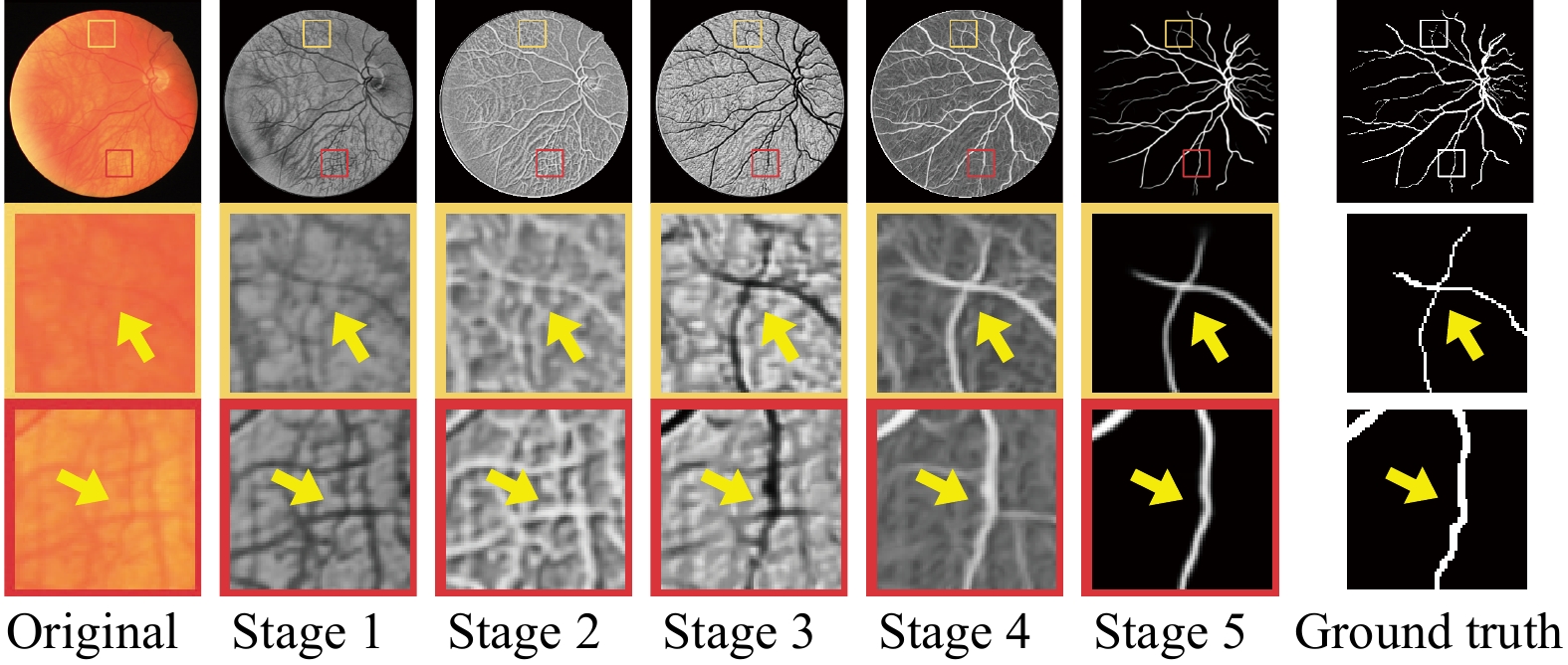
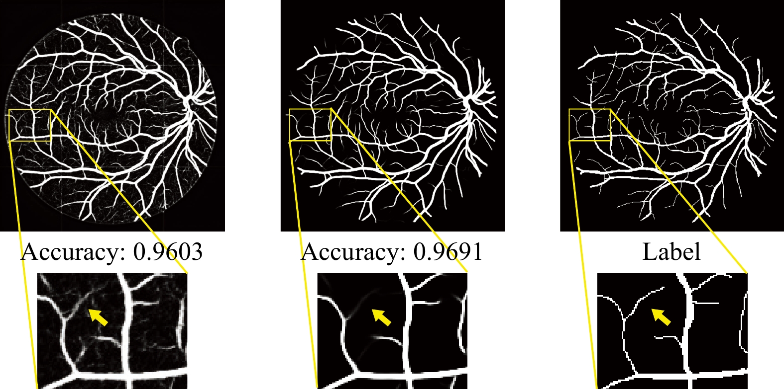
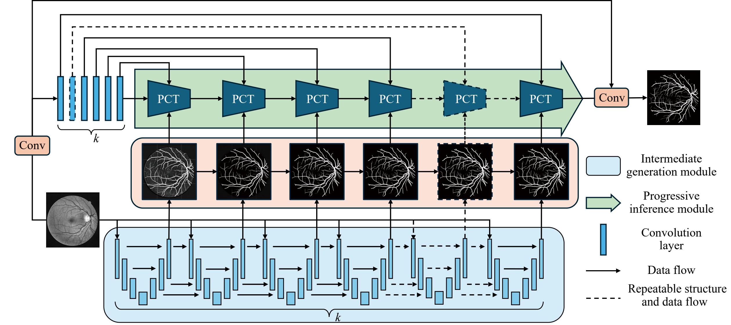
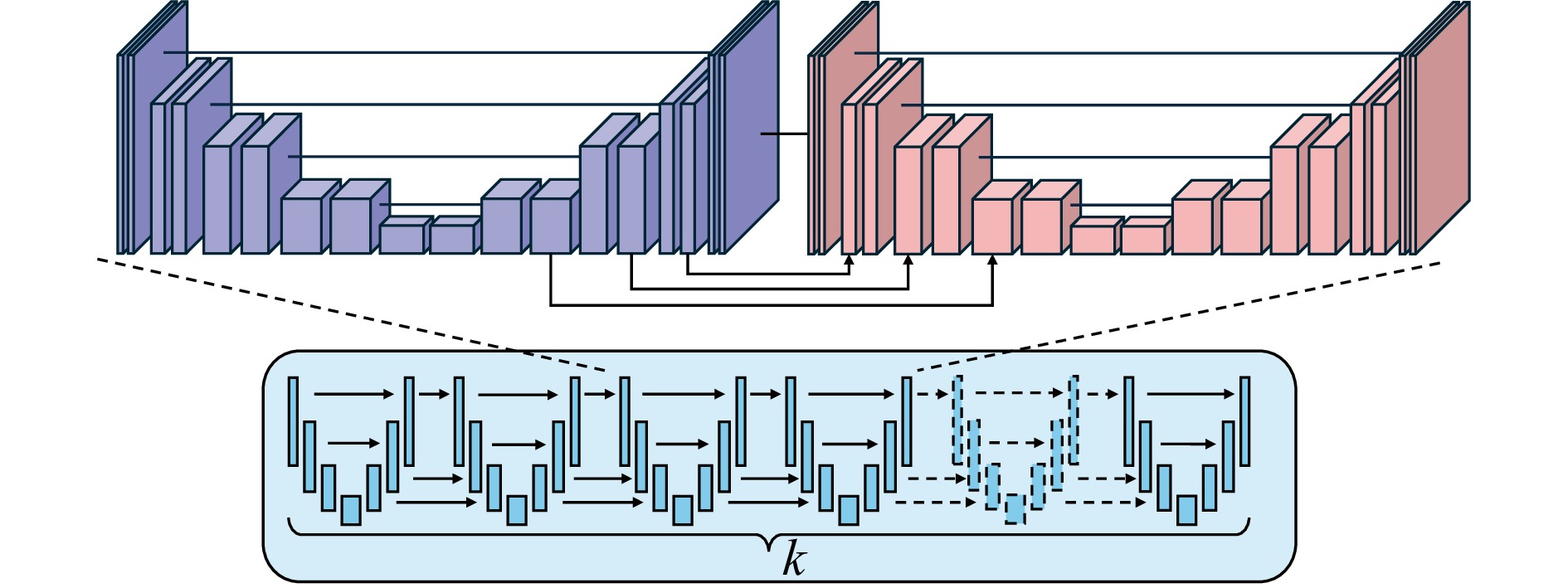
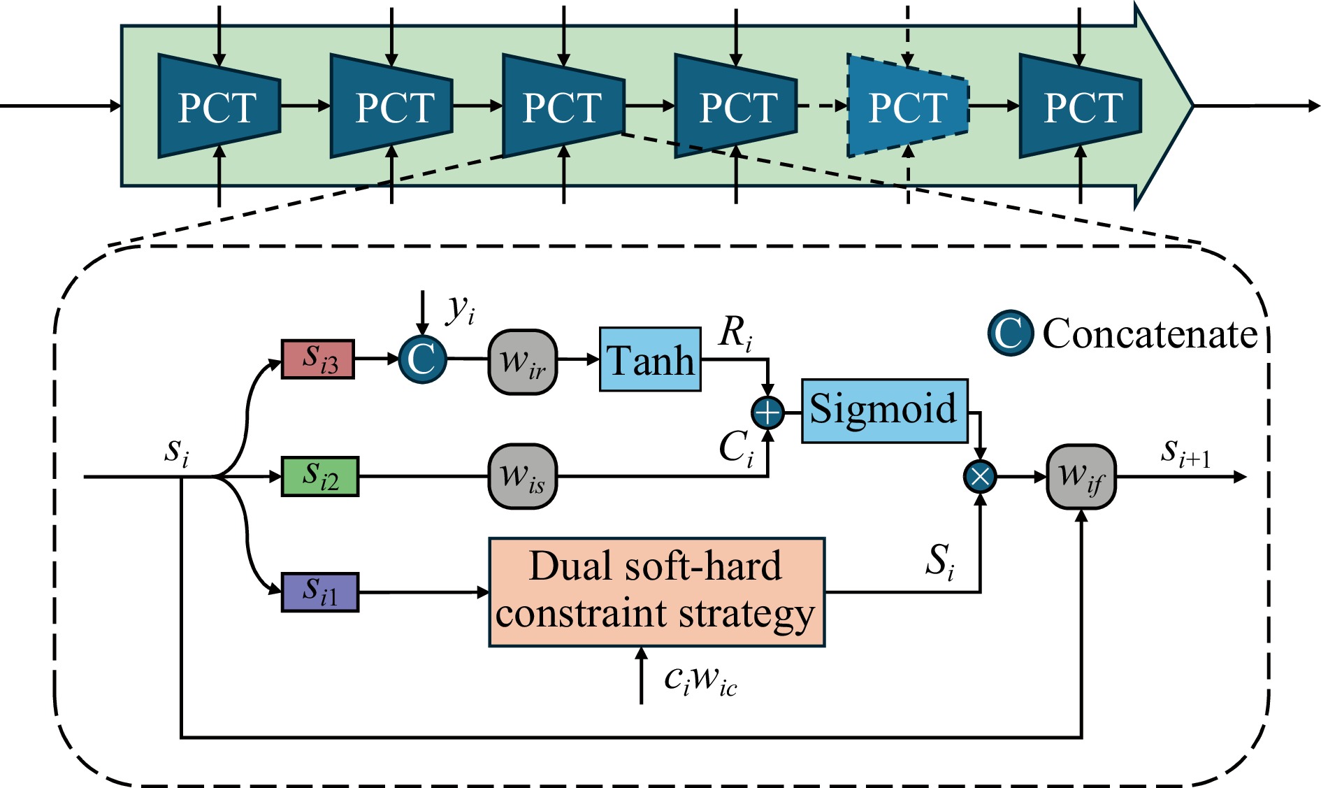
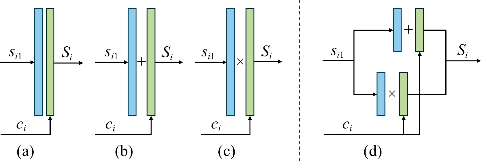
Medical images provide essential information for diagnosing and monitoring various diseases and systemic disorders. With advancements in deep learning and neural networks, numerous methods have been proposed to achieve high-level medical image segmentation results. However, the variability of tiny structures and their high similarity to the background often lead to mis-segmentation in existing methods. To mitigate these challenges, we propose a potential-guided connected network (PCNet) that integrates an innovative dual soft-hard constraint strategy, combining two different progressive supervisions. This strategy modulates the ability of network to differentiate between well-defined and ambiguous structures through a hyper-parameter, thereby enhancing its capability to detect tiny structures. Furthermore, PCNet is composed of two key modules, including the intermediate generation (IG) module and the progressive inference (PI) module. The IG module produces a range of outputs with varying segmentation potentials using a novel serial architecture, which serves as the foundational input for progressive reasoning in the PI module. The PI module, leveraging the outputs of the IG module, is designed to progressively extract comprehensive contextual information, ultimately producing refined segmentation results. PCNet is evaluated on several publicly available datasets, including DRIVE, MoNuSeg, CoNIC, FIVES, and GlaS, achieving accuracy of 96.92%, 90.29%, 93.93%, 98.82%, and 92.00%, respectively. Extensive experiments demonstrate that our model outperforms the current state-of-the-art methods for tiny structure segmentation in medical images.
C. Chen, Y. Song, J. Yi, L. Guo, Z. Lei, and S. Gao, “Potential-guided connected network for tiny structure segmentation in medical images,” IEEE/CAA J. Autom. Sinica, vol. 13, no. 8, pp. 1826–1841, Aug. 2026. doi: 10.1109/JAS.2025.125705.










 E-mail Alert