A journal of IEEE and CAA , publishes
high-quality papers in English on original
theoretical/experimental research
and development in all areas of automation
 Volume 13
Issue 4
Volume 13
Issue 4
IEEE/CAA Journal of Automatica Sinica
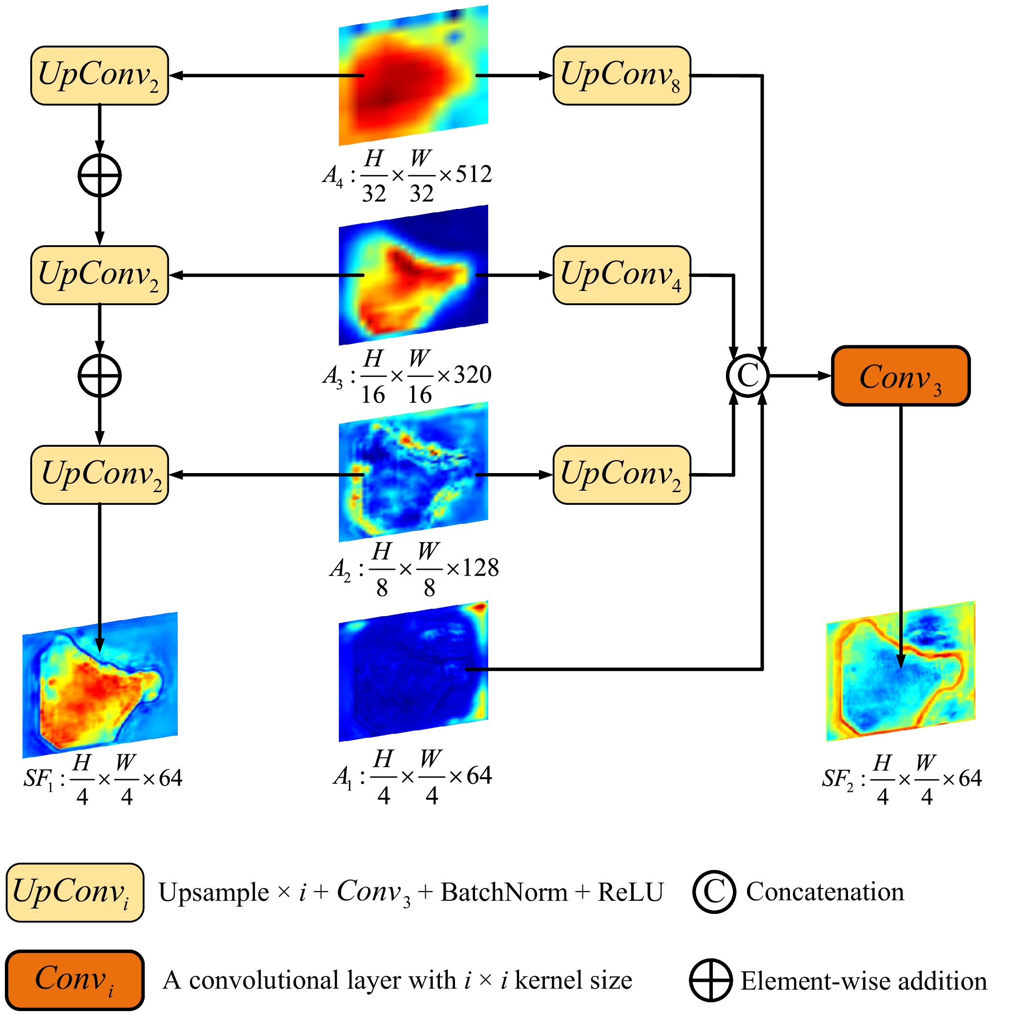
| Citation: | G. Lv, B. Wang, C. Xu, W. Ding, and J. Liu, “MFAINet: Multi-receptive field feature fusion with attention-integrated for polyp segmentation,” IEEE/CAA J. Autom. Sinica, vol. 13, no. 4, pp. 822–836, Apr. 2026. doi: 10.1109/JAS.2025.125408

|

| [1] |
X. Tang, X. Qiao, C. Chen, Y. Liu, J. Zhu, and J. Liu, “Regulation mechanism of long noncoding RNAs in colon cancer development and progression,” Yonsei Med. J., vol. 60, no. 4, pp. 319–325, Mar. 2019. doi: 10.3349/ymj.2019.60.4.319
|
| [2] |
M. P. Singh, S. Rai, A. Pandey, N. K. Singh, and S. Srivastava, “Molecular subtypes of colorectal cancer: An emerging therapeutic opportunity for personalized medicine,” Genes Dis., vol. 8, no. 2, pp. 133–145, Oct. 2019.
|
| [3] |
R. Zhang, G. Li, Z. Li, S. Cui, D. Qian, and Y. Yu, “Adaptive context selection for polyp segmentation,” in Proc. 23rd Int. Conf. Medical Image Computing and Computer Assisted Intervention, Lima, Peru, 2020, pp. 253−262.
|
| [4] |
M. Maida, D. S. Dahiya, Y. R. Shah, A. Tiwari, H. Gopakumar, I. Vohra, A. Khan, F. Jaber, D. Ramai, and A. Facciorusso, “Screening and surveillance of colorectal cancer: A review of the literature,” Cancers, vol. 16, no. 15, Art. no. 2746, Aug. 2024. doi: 10.3390/cancers16152746
|
| [5] |
R. Abedizadeh, F. Majidi, H. R. Khorasani, H. Abedi, and D. Sabour, “Colorectal cancer: A comprehensive review of carcinogenesis, diagnosis, and novel strategies for classified treatments,” Cancer Metastasis Rev., vol. 43, no. 2, pp. 729–753, Jun. 2024. doi: 10.1007/s10555-023-10158-3
|
| [6] |
S. Y. Kim, H.-S. Kim, and H. J. Park, “Adverse events related to colonoscopy: Global trends and future challenges,” World J. Gastroenterol., vol. 25, no. 2, pp. 190–204, Jan. 2019. doi: 10.3748/wjg.v25.i2.190
|
| [7] |
E. Sanderson and B. J. Matuszewski, “FCN-transformer feature fusion for polyp segmentation,” in Proc. 26th Ann. Conf. Medical Image Understanding and Analysis, Cambridge, UK, 2022, pp. 892−907.
|
| [8] |
B. Sushma, C. K. Raghavendra, and J. Prashanth, “CNN based U-Net with modified skip connections for colon polyp segmentation,” in Proc. 5th Int. Conf. Computing Methodologies and Communication, Erode, India, 2021, pp. 1762−1766.
|
| [9] |
D. Jha, P. H. Smedsrud, M. A. Riegler, P. Halvorsen, T. de Lange, D. Johansen, and H. D. Johansen, “Kvasir-SEG: A segmented polyp dataset,” in Proc. 26th Int. Conf. MultiMedia Modeling, Daejeon, South Korea, 2020, pp. 451−462.
|
| [10] |
D. Vázquez, J. Bernal, F. J. Sánchez, G. Fernández-Esparrach, A. M. López, A. Romero, M. Drozdzal, and A. Courville, “A benchmark for endoluminal scene segmentation of colonoscopy images,” J. Healthcare Eng., vol. 2017, Art. no. 4037190, Jul. 2017.
|
| [11] |
J. Silva, A. Histace, O. Romain, X. Dray, and B. Granado, “Toward embedded detection of polyps in WCE images for early diagnosis of colorectal cancer,” Int. J. Comput. Assisted Radiol. Surg., vol. 9, no. 2, pp. 283–293, Mar. 2014. doi: 10.1007/s11548-013-0926-3
|
| [12] |
N. Tajbakhsh, S. R. Gurudu, and J. Liang, “Automated polyp detection in colonoscopy videos using shape and context information,” IEEE Trans. Med. Imaging, vol. 35, no. 2, pp. 630–644, Feb. 2016. doi: 10.1109/TMI.2015.2487997
|
| [13] |
J. Bernal, F. J. Sanchez, G. Fernandez-Esparrach, D. Gil, C. Rodríguez, and F. Vilariño, “WM-DOVA maps for accurate polyp highlighting in colonoscopy: Validation vs. saliency maps from physicians,” Comput. Med. Imaging Graph., vol. 43, pp. 99–111, Jul. 2015. doi: 10.1016/j.compmedimag.2015.02.007
|
| [14] |
O. Ronneberger, P. Fischer, and T. Brox, “U-net: Convolutional networks for biomedical image segmentation,” in Proc. 18th Int. Conf. Medical Image Computing and Computer-Assisted Intervention, Munich, Germany, 2015, pp. 234−241.
|
| [15] |
Z. Zhou, M. M. R. Siddiquee, N. Tajbakhsh, and J. Liang, “UNet++: A nested U-Net architecture for medical image segmentation,” in Proc. 4th Int. Workshop on Deep Learning in Medical Image Analysis and Multimodal Learning for Clinical Decision Support, Granada, Spain, 2018, pp. 3−11.
|
| [16] |
Y. Fang, C. Chen, Y. Yuan, and K.-Y. Tong, “Selective feature aggregation network with area-boundary constraints for polyp segmentation,” in Proc. 22nd Int. Conf. Medical Image Computing and Computer Assisted Intervention, Shenzhen, China, 2019, pp. 302−310.
|
| [17] |
D.-P. Fan, G.-P. Ji, T. Zhou, G. Chen, H. Fu, J. Shen, and L. Shao, “PraNet: Parallel reverse attention network for polyp segmentation,” in Proc. 23rd Int. Conf. Medical Image Computing and Computer Assisted Intervention, Lima, Peru, 2020, pp. 263−273.
|
| [18] |
J. Wei, Y. Hu, R. Zhang, Z. Li, S. K. Zhou, and S. Cui, “Shallow attention network for polyp segmentation,” in Proc. 24th Int. Conf. Medical Image Computing and Computer Assisted Intervention, Strasbourg, France, 2021, pp. 699−708.
|
| [19] |
B. Dong, W. Wang, D.-P. Fan, J. Li, H. Fu, and L. Shao, “Polyp-PVT: Polyp segmentation with pyramid vision transformers,” CAAI Artif. Intell. Res., vol. 2, Art. no. 9150015, 2023. doi: 10.26599/air.2023.9150015
|
| [20] |
N. T. Duc, N. T. Oanh, N. T. Thuy, T. M. Triet, and V. S. Dinh, “ColonFormer: An efficient transformer based method for colon polyp segmentation,” IEEE Access, vol. 10, pp. 80575–80586, Aug. 2022. doi: 10.1109/ACCESS.2022.3195241
|
| [21] |
M. Nguyen, T. T. Bui, Q. Van Nguyen, T. T. Nguyen, and T. Van Pham, “LAPFormer: A light and accurate polyp segmentation transformer,” arXiv preprint arXiv: 2210.04393, 2022.
|
| [22] |
X. Zhao, H. Jia, Y. Pang, L. Lv, F. Tian, L. Zhang, W. Sun, and H. Lu, “M.2SNet: Multi-scale in multi-scale subtraction network for medical image segmentation,” arXiv preprint arXiv: 2303.10894, 2023.
|
| [23] |
J. Liu, Q. Chen, Y. Zhang, Z. Wang, X. Deng, and J. Wang, “Multi-level feature fusion network combining attention mechanisms for polyp segmentation,” Inf. Fusion, vol. 104, Art. no. 102195, Apr. 2024. doi: 10.1016/j.inffus.2023.102195
|
| [24] |
W. Li, Y. Zhao, F. Li, and L. Wang, “MIA-Net: Multi-information aggregation network combining transformers and convolutional feature learning for polyp segmentation,” Knowl. Based Syst., vol. 247, Art. no. 108824, Jul. 2022. doi: 10.1016/j.knosys.2022.108824
|
| [25] |
N.-T. Bui, D.-H. Hoang, Q.-T. Nguyen, M.-T. Tran, and N. Le, “MEGANet: Multi-scale edge-guided attention network for weak boundary polyp segmentation,” in Proc. IEEE/CVF Winter Conf. Applications of Computer Vision, Waikoloa, USA, 2024, pp. 7970−7979.
|
| [26] |
F. Liu, Z. Hua, J. Li, and L. Fan, “DBMF: Dual branch multiscale feature fusion network for polyp segmentation,” Comput. Biol. Med., vol. 151, Art. no. 106304, Dec. 2022. doi: 10.1016/j.compbiomed.2022.106304
|
| [27] |
W. Li, Z. Huang, F. Li, Y. Zhao, and H. Zhang, “CIFG-Net: Cross-level information fusion and guidance network for polyp segmentation,” Comput. Biol. Med., vol. 169, Art. no. 107931, Feb. 2024. doi: 10.1016/j.compbiomed.2024.107931
|
| [28] |
Y. Xia, H. Yun, and Y. Liu, “MFEFNet: Multi-scale feature enhancement and fusion network for polyp segmentation,” Comput. Biol. Med., vol. 157, Art. no. 106735, May 2023. doi: 10.1016/j.compbiomed.2023.106735
|
| [29] |
Z.-U.-D. Muhammad, U. Muhammad, Z. Huang, and N. Gu, “MMFIL-Net: Multi-level and multi-source feature interactive lightweight network for polyp segmentation,” Displays, vol. 81, Art. no. 102600, Jan. 2024. doi: 10.1016/j.displa.2023.102600
|
| [30] |
J. Li, J. Wang, F. Lin, A. Asghar Heidari, Y. Chen, H. Chen, and W. Wu, “PRCNet: A parallel reverse convolutional attention network for colorectal polyp segmentation,” Biomedical Signal Processing and Control, vol 95, Part A, Art. no. 106336, 2024.
|
| [31] |
X. Liu and S. Song, “Attention combined pyramid vision transformer for polyp segmentation,” Biomed. Signal Process. Control, vol. 89, Art. no. 105792, Mar. 2024. doi: 10.1016/j.bspc.2023.105792
|
| [32] |
M. M. Rahman and R. Marculescu, “Medical image segmentation via cascaded attention decoding,” in Proc. IEEE/CVF Winter Conf. Applications of Computer Vision, Waikoloa, USA, 2023, pp. 6211−6220.
|
| [33] |
Ojala T and Pietikäinen M, “Unsupervised texture segmentation using feature distributions,” Pattern Recognition, vol. 32, no. 3, pp. 477–486, 1999.
|
| [34] |
A. V. Mamonov, I. N. Figueiredo, P. N. Figueiredo, and Y.-H. R. Tsai, “Automated polyp detection in colon capsule endoscopy,” IEEE Trans. Med. Imaging, vol. 33, no. 7, pp. 1488–1502, Jul. 2014. doi: 10.1109/TMI.2014.2314959
|
| [35] |
S. Y. Park, D. Sargent, I. Spofford, K. G. Vosburgh, and Y. A. Rahim, “A colon video analysis framework for polyp detection,” IEEE Trans. Biomed. Eng., vol. 59, no. 5, pp. 1408–1418, May 2012. doi: 10.1109/TBME.2012.2188397
|
| [36] |
C. Van Wijk, V. F. Van Ravesteijn, F. M. Vos, and L. J. Van Vliet, “Detection and segmentation of colonic polyps on implicit isosurfaces by second principal curvature flow,” IEEE Trans. Med. Imaging, vol. 29, no. 3, pp. 688–698, Mar. 2010. doi: 10.1109/TMI.2009.2031323
|
| [37] |
D. Jha, P. H. Smedsrud, M. A. Riegler, D. Johansen, T. de Lange, P. Halvorsen, and H. D. Johansen, “ResUNet++: An advanced architecture for medical image segmentation,” in Proc. IEEE Int. Symp. Multimedia, San Diego, USA, 2019, pp. 225−2255.
|
| [38] |
T. Zhou, Y. Zhou, K. He, C. Gong, J. Yang, H. Fu, and D. Shen, “Cross-level feature aggregation network for polyp segmentation,” Pattern Recognit., vol. 140, Art. no. 109555, Aug. 2023. doi: 10.1016/j.patcog.2023.109555
|
| [39] |
B. Murugesan, K. Sarveswaran, S. M. Shankaranarayana, K. Ram, J. Joseph, and M. Sivaprakasam, “Psi-Net: Shape and boundary aware joint multi-task deep network for medical image segmentation,” in Proc. 41st Ann. Int. Conf. IEEE Engineering in Medicine and Biology Society, Berlin, Germany, 2019, pp. 7223−7226.
|
| [40] |
Y. Zhang, H. Liu, and Q. Hu, “Transfuse: Fusing transformers and CNNs for medical image segmentation,” in Proc. 24th Int. Conf. Medical Image Computing and Computer Assisted Intervention, Strasbourg, France, 2021, pp. 14−24.
|
| [41] |
W. Wang, E. Xie, X. Li, D.-P. Fan, K. Song, D. Liang, T. Lu, P. Luo, and L. Shao, “PVT v2: Improved baselines with pyramid vision transformer,” Comput. Visual Med., vol. 8, no. 3, pp. 415–424, Sep. 2022. doi: 10.1007/s41095-022-0274-8
|
| [42] |
R. Margolin, L. Zelnik-Manor, and A. Tal, “How to evaluate foreground maps?” in Proc. IEEE Conf. Computer Vision and Pattern Recognition, Columbus, USA, 2014, pp. 248−255.
|
| [43] |
F. Perazzi, P. Krahenbuhl, Y. Pritch, and A. Hornung, “Saliency filters: Contrast based filtering for salient region detection,” in Proc. IEEE Conf. Computer Vision and Pattern Recognition, Providence, USA, 2012, pp. 733−740.
|
| [44] |
I. Loshchilov and F. Hutter, “Decoupled weight decay regularization,” in Proc. 7th Int. Conf. Learning Representations, New Orleans, USA, 2019.
|
| [45] |
J. Wei, S. Wang, and Q. Huang, “F.3Net: Fusion, feedback and focus for salient object detection,” in Proc. 34th AAAI Conf. Artificial Intelligence, New York, USA, 2020, pp. 12321−12328.
|
| [46] |
C.-H. Huang, H.-Y. Wu, and Y.-L. Lin, “HarDNet-MSEG: A simple encoder-decoder polyp segmentation neural network that achieves over 0.9 mean dice and 86 FPS,” arXiv preprint arXiv: 2101.07172, 2021.
|
| [47] |
K. Patel, A. M. Bur, and G. Wang, “Enhanced U-Net: A feature enhancement network for polyp segmentation,” in Proc. 18th Conf. Robots and Vision, Burnaby, Canada, 2021, pp. 181−188.
|
| [48] |
P. Naylor, M. Laé, F. Reyal, and T. Walter, “Segmentation of nuclei in histopathology images by deep regression of the distance map,” IEEE Trans. Med. Imaging, vol. 38, no. 2, pp. 448–459, Feb. 2019. doi: 10.1109/TMI.2018.2865709
|
| [49] |
R. Azad, M. Asadi-Aghbolaghi, M. Fathy, and S. Escalera, “Bi-directional convLSTM U-Net with densley connected convolutions,” in Proc. IEEE/CVF Int. Conf. Computer Vision Workshop, Seoul, Korea (South), 2019, pp. 406−415.
|
Figures(12) / Tables(4)


 DownLoad: